Contents Yves Clermont, Michael Lalli, Zsuzsanna Bencsath-Makkai
LIGHT-MICROSCOPIC HISTOLOGY ATLAS
     11. Oral Cavity
  1     2     3     4     5     6     7     8     9     10     11     12     13     14     15     16     17     18     19     20     21     22     23     24     25  
  26     27     28     29     30     31     32     33     34     35     36     37     38     39     40     41  

    11.39   Tooth formation Version française
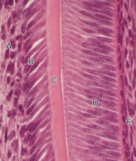
Figures 11.39, 11.40 and 11.41 were taken along the continuously growing root of a rodent’s incisor.

This image was taken close to the tip of the root. It shows the following layers or components (left to right):

  1. Connective tissue of the pulp
  2. Tall secretory odontoblasts
  3. Thin layer of dentin
  4. Tall non-secretory ameloblasts
  5. Intermediate layer, or stratum intermedium, of the enamel organ (see Figure 11.33)

No enamel is seen at this early stage of tooth growth.

Stain: H–­­E
Magnification: ×600


We'd very much appreciate receiving your comments and suggestions!We'd very much appreciate receiving your comments and suggestions!

Creative Commons Licence The text and images of this Histology Atlas, by Yves Clermont, Michael Lalli & Zsuzsanna Bencsath-Makkai, are licensed under a Creative Commons Attribution-Noncommercial-No Derivative Works 2.5 Canada Licence and cannot be modified without the written permission of the authors. Use of any text or images must carry an acknowledgement which includes a link to the original work.


HAweb/Zs B-M -> HAbase-generated on 2013-09-13