Contents Yves Clermont, Michael Lalli, Zsuzsanna Bencsath-Makkai
LIGHT-MICROSCOPIC HISTOLOGY ATLAS
     11. Oral Cavity
  1     2     3     4     5     6     7     8     9     10     11     12     13     14     15     16     17     18     19     20     21     22     23     24     25  
  26     27     28     29     30     31     32     33     34     35     36     37     38     39     40     41  

    11.5   Tongue Version française
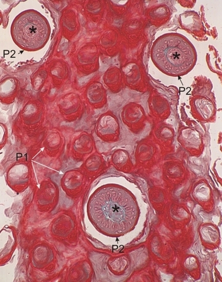
Section of the tongue of a bat. The plane of section is tangential to the upper surface of the tongue.

This field shows many transversely cut filiform papillae (P1) and three transverse sections of fungiform papillae (P2).

The large fungiform papilla (below) is surrounded by a deep furrow. These fungiform papillae show a core of connective tissue (*) surrounded by a stratified squamous epithelium.

The keratin of the filiform and fungiform papillae is stained bright red with Masson’s trichrome.

Stain: Masson’s Trichrome
Magnification: ×150


We'd very much appreciate receiving your comments and suggestions!We'd very much appreciate receiving your comments and suggestions!

Creative Commons Licence The text and images of this Histology Atlas, by Yves Clermont, Michael Lalli & Zsuzsanna Bencsath-Makkai, are licensed under a Creative Commons Attribution-Noncommercial-No Derivative Works 2.5 Canada Licence and cannot be modified without the written permission of the authors. Use of any text or images must carry an acknowledgement which includes a link to the original work.


HAweb/Zs B-M -> HAbase-generated on 2013-09-13