Contents Yves Clermont, Michael Lalli, Zsuzsanna Bencsath-Makkai
LIGHT-MICROSCOPIC HISTOLOGY ATLAS
     11. Oral Cavity
  1     2     3     4     5     6     7     8     9     10     11     12     13     14     15     16     17     18     19     20     21     22     23     24     25  
  26     27     28     29     30     31     32     33     34     35     36     37     38     39     40     41  

    11.37   Tooth formation Version française
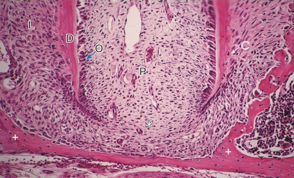
Tip of a root of a growing molar of a rodent.

This field shows the pulp cavity (P) and the apical foramen (*), or pore at the tip of the root. The connective tissue of the pulp is continuous with the periodontal connective tissue.

The inside wall of the root shows the odontoblasts (O) applied to the newly formed dentin (D).

Some cementocytes (C) are visible on the external surface of the dentin.

The connective tissue of the periodontal ligament (L) and the bone tissue (+) are also labelled.

Stain: H–­­E
Magnification: ×300


We'd very much appreciate receiving your comments and suggestions!We'd very much appreciate receiving your comments and suggestions!

Creative Commons Licence The text and images of this Histology Atlas, by Yves Clermont, Michael Lalli & Zsuzsanna Bencsath-Makkai, are licensed under a Creative Commons Attribution-Noncommercial-No Derivative Works 2.5 Canada Licence and cannot be modified without the written permission of the authors. Use of any text or images must carry an acknowledgement which includes a link to the original work.


HAweb/Zs B-M -> HAbase-generated on 2013-09-13