Contents Yves Clermont, Michael Lalli, Zsuzsanna Bencsath-Makkai
LIGHT-MICROSCOPIC HISTOLOGY ATLAS
     11. Oral Cavity
  1     2     3     4     5     6     7     8     9     10     11     12     13     14     15     16     17     18     19     20     21     22     23     24     25  
  26     27     28     29     30     31     32     33     34     35     36     37     38     39     40     41  
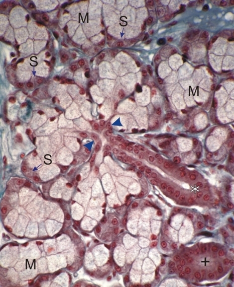
    11.17   Salivary gland Version française
A sublingual gland.

This field shows the difference between the pale mucous cells (M) and the deeply stained serous demilune cells (S).

It also shows several mixed acini radiating from a short intercalated duct (arrowheads). The intercalated duct is continuous with a more acidophilic striated duct (*). Another striated duct (+) cut obliquely is also visible (bottom right).

A small amount of connective tissue stained green is seen between the acini.

Stain: Masson’s Trichrome
Magnification: ×800


We'd very much appreciate receiving your comments and suggestions!We'd very much appreciate receiving your comments and suggestions!

Creative Commons Licence The text and images of this Histology Atlas, by Yves Clermont, Michael Lalli & Zsuzsanna Bencsath-Makkai, are licensed under a Creative Commons Attribution-Noncommercial-No Derivative Works 2.5 Canada Licence and cannot be modified without the written permission of the authors. Use of any text or images must carry an acknowledgement which includes a link to the original work.


HAweb/Zs B-M -> HAbase-generated on 2013-09-13