Contents Yves Clermont, Michael Lalli, Zsuzsanna Bencsath-Makkai
LIGHT-MICROSCOPIC HISTOLOGY ATLAS
     11. Oral Cavity
  1     2     3     4     5     6     7     8     9     10     11     12     13     14     15     16     17     18     19     20     21     22     23     24     25  
  26     27     28     29     30     31     32     33     34     35     36     37     38     39     40     41  

    11.34   Tooth formation Version française
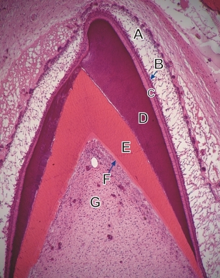
Section of a growing incisor.

This field shows the developing crown, with a progressive increase in thickness of the enamel and dentin.

Compare with the drawing in Figure 11.33.

The following structures are labelled (top to bottom):

  1. Stellate reticulum of the enamel organ
  2. Intermediate layer
  3. Layer of ameloblasts
  4. Basophilic enamel
  5. Acidophilic dentin
  6. Layer of odontoblasts
  7. Dental pulp

Stain: H–­­E
Magnification: ×150


We'd very much appreciate receiving your comments and suggestions!We'd very much appreciate receiving your comments and suggestions!

Creative Commons Licence The text and images of this Histology Atlas, by Yves Clermont, Michael Lalli & Zsuzsanna Bencsath-Makkai, are licensed under a Creative Commons Attribution-Noncommercial-No Derivative Works 2.5 Canada Licence and cannot be modified without the written permission of the authors. Use of any text or images must carry an acknowledgement which includes a link to the original work.


HAweb/Zs B-M -> HAbase-generated on 2013-09-13