Contents Yves Clermont, Michael Lalli, Zsuzsanna Bencsath-Makkai
LIGHT-MICROSCOPIC HISTOLOGY ATLAS
     11. Oral Cavity
  1     2     3     4     5     6     7     8     9     10     11     12     13     14     15     16     17     18     19     20     21     22     23     24     25  
  26     27     28     29     30     31     32     33     34     35     36     37     38     39     40     41  

    11.27   Tooth Version française
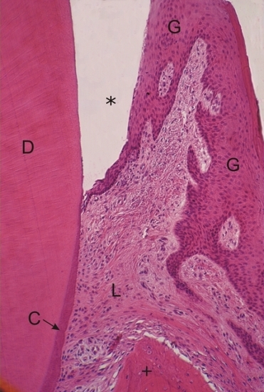
Section of a tooth (left) and of the gingiva (right).

The enamel (*), which has been dissolved by a chelating agent, is located between the acidophilic dentin (D) and the epithelium of the gingiva (G).

The acellular cementum (C) is a thin basophilic layer at the surface of the dentin of the root.

The dense connective tissue of the periodontal ligament (L) and bone (+) are also indicated.

Stain: H–­­E
Magnification: ×240


We'd very much appreciate receiving your comments and suggestions!We'd very much appreciate receiving your comments and suggestions!

Creative Commons Licence The text and images of this Histology Atlas, by Yves Clermont, Michael Lalli & Zsuzsanna Bencsath-Makkai, are licensed under a Creative Commons Attribution-Noncommercial-No Derivative Works 2.5 Canada Licence and cannot be modified without the written permission of the authors. Use of any text or images must carry an acknowledgement which includes a link to the original work.


HAweb/Zs B-M -> HAbase-generated on 2013-09-13