Contents Yves Clermont, Michael Lalli, Zsuzsanna Bencsath-Makkai
LIGHT-MICROSCOPIC HISTOLOGY ATLAS
     11. Oral Cavity
  1     2     3     4     5     6     7     8     9     10     11     12     13     14     15     16     17     18     19     20     21     22     23     24     25  
  26     27     28     29     30     31     32     33     34     35     36     37     38     39     40     41  

    11.26   Tooth Version française
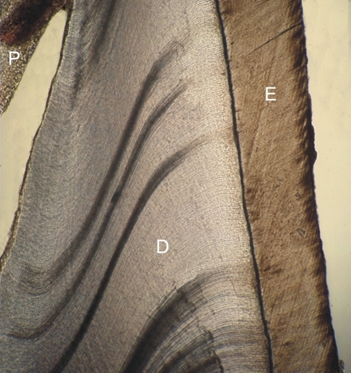
Thin slice of an undecalcified human incisor.

This unstained section of the crown shows the enamel (E) and the dentin (D) next to the retracted pulp (P).

The enamel, 96% mineralized, shows a number of striations. The dentin, 70% mineralized, shows fine wavy dentinal tubules running from the enamel to the pulp (P).

The dentinal tubules contain fine cytoplasmic processes of odontoblasts which are responsible for the formation of the dentin.

Stain: None
Magnification: ×10


We'd very much appreciate receiving your comments and suggestions!We'd very much appreciate receiving your comments and suggestions!

Creative Commons Licence The text and images of this Histology Atlas, by Yves Clermont, Michael Lalli & Zsuzsanna Bencsath-Makkai, are licensed under a Creative Commons Attribution-Noncommercial-No Derivative Works 2.5 Canada Licence and cannot be modified without the written permission of the authors. Use of any text or images must carry an acknowledgement which includes a link to the original work.


HAweb/Zs B-M -> HAbase-generated on 2013-09-13