|
||
| 11. Oral Cavity | ||
| 1 2 3 4 5 6 7 8 9 10 11 12 13 14 15 16 17 18 19 20 21 22 23 24 25 | ||
| 26 27 28 29 30 31 32 33 34 35 36 37 38 39 40 41 |
| |||
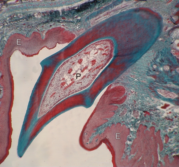 |
Oblique section of the incisor of a bat.
This section shows the dentin of the root (top) and of the crown (bottom) stained green and red. The enamel capping the crown was dissolved during the histological procedure. The well-vascularized pulp (P) occupies the centre of the tooth. The root canal with its apical foramen is not included in this oblique section. The connective tissue attaching the root of the tooth to the bone is green (*). The soft keratin stains deep red at the surface of the gingival epithelium (E). Stain: Massons Trichrome
|
||