|
||
| 11. Oral Cavity | ||
| 1 2 3 4 5 6 7 8 9 10 11 12 13 14 15 16 17 18 19 20 21 22 23 24 25 | ||
| 26 27 28 29 30 31 32 33 34 35 36 37 38 39 40 41 |
| |||
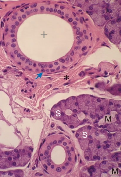 |
Submandibular gland of a dog.
This field shows serous acini (S) and occasional mucous cells (M) in some of them. In proximity to the lobule in an interlobular septum (*) is a large interlobular duct (+). This duct is lined with an epithelium composed of columnar cells and several basal cells (arrow) sitting on the basement membrane. Stain: HE
|
||