Contents Yves Clermont, Michael Lalli, Zsuzsanna Bencsath-Makkai
LIGHT-MICROSCOPIC HISTOLOGY ATLAS
     11. Oral Cavity
  1     2     3     4     5     6     7     8     9     10     11     12     13     14     15     16     17     18     19     20     21     22     23     24     25  
  26     27     28     29     30     31     32     33     34     35     36     37     38     39     40     41  
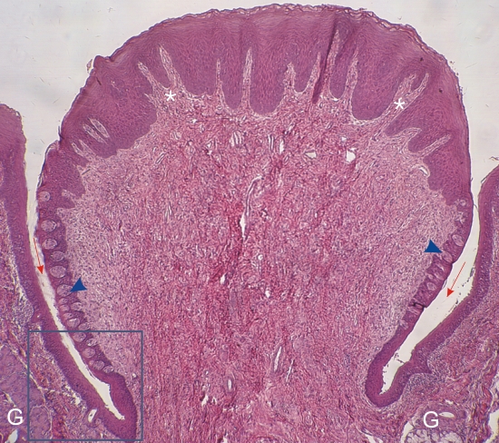
    11.6   Circumvallate papilla Version française
Section of a cat’s tongue. The framed area is magnified in Figure 11.7.

This field shows a circumvallate papilla, one of a few large papillae forming a V-line on the posterior surface of the tongue. This button-shaped papilla is surrounded by a deep circular furrow, or crypt (red arrows).

Numerous taste buds (arrowheads) are seen along the wall of the papilla.

The thick stratified squamous epithelium at the apex of the papilla is deeply invaginated by fingerlike connective tissue projections (*).

Several serous glands of Von Ebner (G) open at the bottom of the crypt.

Stain: H–­­E
Magnification: ×100


We'd very much appreciate receiving your comments and suggestions!We'd very much appreciate receiving your comments and suggestions!

Creative Commons Licence The text and images of this Histology Atlas, by Yves Clermont, Michael Lalli & Zsuzsanna Bencsath-Makkai, are licensed under a Creative Commons Attribution-Noncommercial-No Derivative Works 2.5 Canada Licence and cannot be modified without the written permission of the authors. Use of any text or images must carry an acknowledgement which includes a link to the original work.


HAweb/Zs B-M -> HAbase-generated on 2013-09-13