Sommaire Yves Clermont, Michael Lalli, Zsuzsanna Bencsath-Makkai
ATLAS D'HISTOLOGIE EN MICROSCOPIE OPTIQUE
     11. Cavité Orale
  1     2     3     4     5     6     7     8     9     10     11     12     13     14     15     16     17     18     19     20     21     22     23     24     25  
  26     27     28     29     30     31     32     33     34     35     36     37     38     39     40     41  
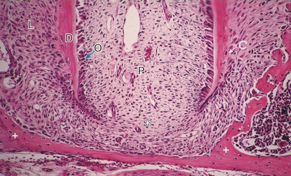
    11.37   Cavité buccale: formation de la dent English version
L’extrémité d’une racine de molaire en croissance d’un rongeur.

Ce champ montre la pulpe de la dent (P) et le pore apical à l’extrémité du canal dentaire (*). Le tissu conjonctif de la pulpe est continu avec le tissu conjonctif périodontal.

Sur la face interne de la racine on note la rangée d’odontoblastes (O) le long de la dentine (D) nouvellement formée.

Quelques cémentocytes (C) sont visibles à la surface de la dentine.

Le tissu conjonctif du ligament périodontal (L) et le tissu osseux environnant (+) sont également identifiés.

Coloration: H–­­É
Grossissement: ×300


We'd very much appreciate receiving your comments and suggestions!We'd very much appreciate receiving your comments and suggestions!

Creative Commons Licence The text and images of this Histology Atlas, by Yves Clermont, Michael Lalli & Zsuzsanna Bencsath-Makkai, are licensed under a Creative Commons Attribution-Noncommercial-No Derivative Works 2.5 Canada Licence and cannot be modified without the written permission of the authors. Use of any text or images must carry an acknowledgement which includes a link to the original work.


HAweb/Zs B-M -> HAbase-generated on 2013-09-13