Sommaire Yves Clermont, Michael Lalli, Zsuzsanna Bencsath-Makkai
ATLAS D'HISTOLOGIE EN MICROSCOPIE OPTIQUE
     11. Cavité Orale
  1     2     3     4     5     6     7     8     9     10     11     12     13     14     15     16     17     18     19     20     21     22     23     24     25  
  26     27     28     29     30     31     32     33     34     35     36     37     38     39     40     41  
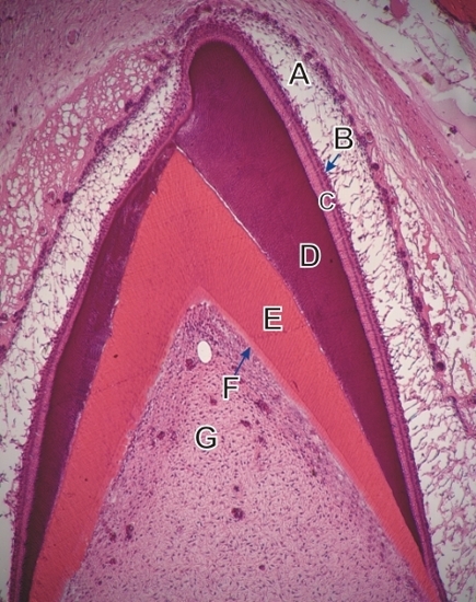
    11.34   Cavité buccale: formation de la dent English version
Coupe d’une incisive en croissance d’un singe. Ce champ, qui correspond aux schémas de la figure 11.33, montre la couronne de la dent en formation et l’épaississement progressif de la dentine et de l’émaill.

Les structures suivantes sont identifiées (du haut vers le bas):

  1. Réticulum étoilé de l’organe de l’émail
  2. Couche épithéliale intermédiaire
  3. Couche des améloblastes
  4. L’émail basophile
  5. Dentine acidophile
  6. Couche d’odontoblastes
  7. Pulpe dentaire

Coloration: H–­­É
Grossissement: ×150


We'd very much appreciate receiving your comments and suggestions!We'd very much appreciate receiving your comments and suggestions!

Creative Commons Licence The text and images of this Histology Atlas, by Yves Clermont, Michael Lalli & Zsuzsanna Bencsath-Makkai, are licensed under a Creative Commons Attribution-Noncommercial-No Derivative Works 2.5 Canada Licence and cannot be modified without the written permission of the authors. Use of any text or images must carry an acknowledgement which includes a link to the original work.


HAweb/Zs B-M -> HAbase-generated on 2013-09-13