Sommaire Yves Clermont, Michael Lalli, Zsuzsanna Bencsath-Makkai
ATLAS D'HISTOLOGIE EN MICROSCOPIE OPTIQUE
     11. Cavité Orale
  1     2     3     4     5     6     7     8     9     10     11     12     13     14     15     16     17     18     19     20     21     22     23     24     25  
  26     27     28     29     30     31     32     33     34     35     36     37     38     39     40     41  
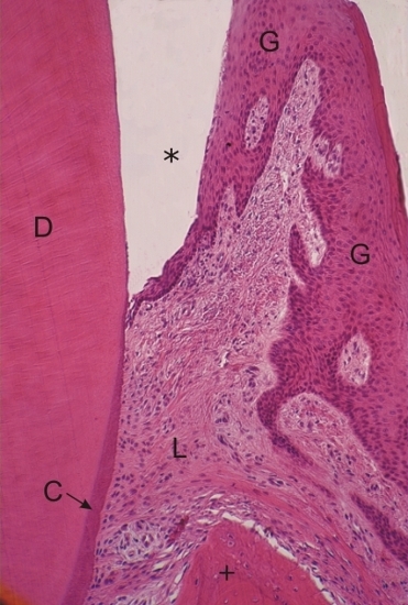
    11.27   Cavité buccale: la dent English version
Coupe d’une coupe de dent à gauche et de la gencive à droite.

L’émail (*) de la couronne, dissout au cours de la préparation de la coupe, est logé dans l’espace entre la dentine (D) et l’épithélium de la gencive (G).

Le cément acellulaire (C) est une couche basophile à la surface de la dentine de la racine.

Le tissu conjonctif du ligament périodontal (L) ainsi que l’os (+) sont également identifiés.

Coloration: H–­­É
Grossissement: ×240


We'd very much appreciate receiving your comments and suggestions!We'd very much appreciate receiving your comments and suggestions!

Creative Commons Licence The text and images of this Histology Atlas, by Yves Clermont, Michael Lalli & Zsuzsanna Bencsath-Makkai, are licensed under a Creative Commons Attribution-Noncommercial-No Derivative Works 2.5 Canada Licence and cannot be modified without the written permission of the authors. Use of any text or images must carry an acknowledgement which includes a link to the original work.


HAweb/Zs B-M -> HAbase-generated on 2013-09-13