|
||
| 1. Epithelia | ||
| 1 2 3 4 5 6 7 8 9 10 11 12 13 14 15 16 17 18 19 20 21 22 23 24 |
| |||
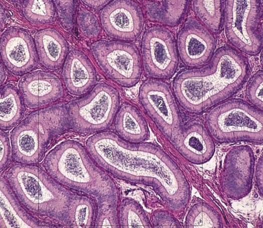 |
Higher-power view of the framed area in Figure 1.1B (×70).
The epididymis is formed of a single coiled duct seen here in transverse (1), oblique (2) and longitudinal (3) sections. The duct is lined with epithelium, the details of which are still unclear at this magnification. The intensely stained material in the centre of the duct is composed of clustered spermatozoa. The framed area is magnified 200 times in Figure 1.3. Stain: HE
|
||