|
||
| 1. Epithelia | ||
| 1 2 3 4 5 6 7 8 9 10 11 12 13 14 15 16 17 18 19 20 21 22 23 24 |
| |||
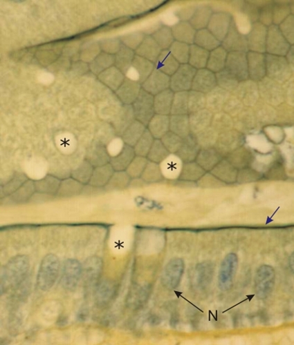 |
Simple columnar epithelium of intestinal villi of a rodent.
This section, treated with TPA, shows intense green staining of the terminal bar-terminal web complexes (arrows) at the apexes of the columnar cells. In the field below, the plane of section is perpendicular to the surface of the epithelium. At the apex the terminal bar-terminal web complexes are seen sideways (the overlying striated border is not stained). The nuclei of columnar cells (N) are lightly stained. In the field above, the plane of section is tangential and parallel to the epithelial surface. The terminal bars clearly delimit the polyhedral boundaries of individual columnar cells. The unstained white profiles (*) in the epithelia of the fields above and below correspond to the mucous secretory pole of goblet cells (see these cells in Figure 1.14). Stain: TPA
|
||