|
||
| 1. Epithelia | ||
| 1 2 3 4 5 6 7 8 9 10 11 12 13 14 15 16 17 18 19 20 21 22 23 24 |
| |||
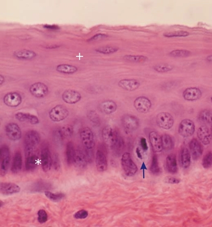 |
Stratified squamous epithelium (not keratinized) of the oral cavity of a rodent.
This image shows several layers of epithelial cells but their borderlines are barely visible in this section. In the basal cells the vertical nuclei are heavily stained (*). The arrow points to a mitotic figure. The cells, derived from the basal cells, become polyhedral and migrate toward the surface of the epithelium where they become squamous (+) and eventually desquamate. Stain: HE
|
||