Contents Yves Clermont, Michael Lalli, Zsuzsanna Bencsath-Makkai
LIGHT-MICROSCOPIC HISTOLOGY ATLAS
     1. Epithelia
  1     2     3     4     5     6     7     8     9     10     11     12     13     14     15     16     17     18     19     20     21     22     23     24  

    1.12   Simple columnar epithelium of the small intestine Version française
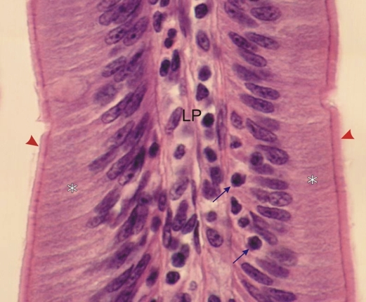
Small portion of an intestinal villus lined with a simple columnar epithelium.

The cytoplasm of the epithelial cells (*) is long, slender and here slightly curved, with an elongated nucleus at its base. At the apexes of these cells, the acidophilic terminal bar-terminal web complex underlies the striated border which is composed of microvilli (arrowheads).

The epithelium is invaded by lymphocytes (arrows) coming from the subjacent connective tissue or lamina propria (LP).

Stain: H–­­E
Magnification: ×700


We'd very much appreciate receiving your comments and suggestions!We'd very much appreciate receiving your comments and suggestions!

Creative Commons Licence The text and images of this Histology Atlas, by Yves Clermont, Michael Lalli & Zsuzsanna Bencsath-Makkai, are licensed under a Creative Commons Attribution-Noncommercial-No Derivative Works 2.5 Canada Licence and cannot be modified without the written permission of the authors. Use of any text or images must carry an acknowledgement which includes a link to the original work.


HAweb/Zs B-M -> HAbase-generated on 2013-09-13