|
||
| 1. Epithelia | ||
| 1 2 3 4 5 6 7 8 9 10 11 12 13 14 15 16 17 18 19 20 21 22 23 24 |
| |||
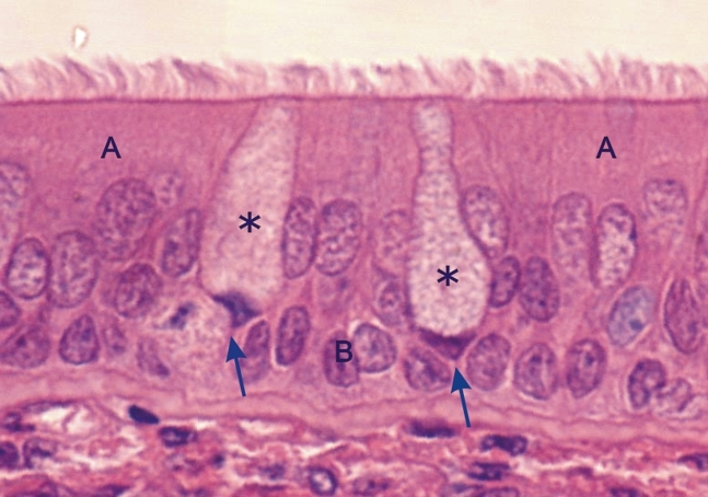 |
Pseudostratified columnar ciliated epithelium of the mucosa of a dog trachea.
This field shows, in addition to basal cells (B), two types of columnar epithelial cells, i.e., tall columnar ciliated cells (A) and goblet cells (*). The goblet cells show dark triangular nuclei (arrows) under a large pocket of cytoplasm containing mucigen granules, the precursors of mucus. Stain: HE
|
||