Contents Yves Clermont, Michael Lalli, Zsuzsanna Bencsath-Makkai
LIGHT-MICROSCOPIC HISTOLOGY ATLAS
     1. Epithelia
  1     2     3     4     5     6     7     8     9     10     11     12     13     14     15     16     17     18     19     20     21     22     23     24  

    1.14   Intestinal villi cut transversely and stained with PA-Schiff to show glycoproteins Version française
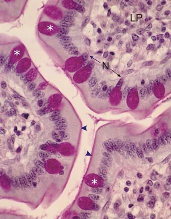
Transverse sections of intestinal villi of a dog stained with periodic acid-Schiff (PAS) and hematoxylin. PAS stains glycoproteins purple.

In this section, PAS stains the mucigen granules of the goblet cells (*). These goblet cells are intercalated between the other pale columnar epithelial cells that compose this simple columnar epithelium.

The striated borders of these columnar cells are also PAS-positive owing to the carbohydrate-rich cell coat (glycocalyx) associated with the membrane of the microvilli (arrowheads).

The nuclei of goblet cells (N) and the lamina propria (LP) underlying the epithelium are also indicated.

Stain: PAS-Hematoxylin
Magnification: ×700


We'd very much appreciate receiving your comments and suggestions!We'd very much appreciate receiving your comments and suggestions!

Creative Commons Licence The text and images of this Histology Atlas, by Yves Clermont, Michael Lalli & Zsuzsanna Bencsath-Makkai, are licensed under a Creative Commons Attribution-Noncommercial-No Derivative Works 2.5 Canada Licence and cannot be modified without the written permission of the authors. Use of any text or images must carry an acknowledgement which includes a link to the original work.


HAweb/Zs B-M -> HAbase-generated on 2013-09-13