|
||
| 1. Epithelia | ||
| 1 2 3 4 5 6 7 8 9 10 11 12 13 14 15 16 17 18 19 20 21 22 23 24 |
| |||
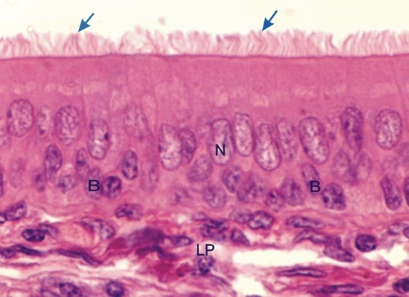 |
Epithelium lining the tracheal mucosa of a dog.
This epithelium shows two layers of nuclei. The nuclei of the basal epithelial cells (B) are small and their cytoplasms are closely applied to the connective tissue. The overlying epithelial columnar cells have large and pale ovoid nuclei (N). They have motile cilia at their luminal surface (arrows). The pointed infranuclear cytoplasm of these cells, (not visible in this light-microscope image), extends between the basal cells and reaches the connective tissue. The term pseudostratified is used to characterize this epithelium to indicate that although the epithelium looks stratified, in fact, all epithelial cells are connected to the connective tissue of the lamina propria (LP). Stain: HE
|
||