|
||
| 1. Epithelia | ||
| 1 2 3 4 5 6 7 8 9 10 11 12 13 14 15 16 17 18 19 20 21 22 23 24 |
| |||
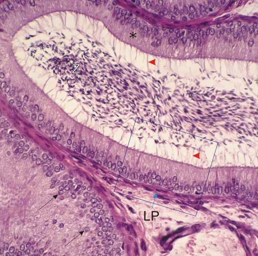 |
Higher-power view (×300) showing details of the simple columnar epithelium of the epididymal duct.
Top, centre: The plane of cut is perpendicular to the epithelium (*), which is 100 µm thick. The long filamentous projections (red arrowheads) at the apex of the epithelial cells correspond to non-motile bundles of submicroscopic microvilli. These bundles are also called stereocilia. Top, left: In the centre of the lumen of the duct, the basophilic small nuclei of spermatozoa (Z) are clearly seen. Bottom, left: The epithelium of the duct is cut tangentially or obliquely and part of the section traverses the layer of nuclei (arrows). The epithelium of the duct is sitting on a connective tissue layer or lamina propria (LP). The framed area is magnified 800 times in Figure 1.5. Stain: HE
|
||