Sommaire Yves Clermont, Michael Lalli, Zsuzsanna Bencsath-Makkai
ATLAS D'HISTOLOGIE EN MICROSCOPIE OPTIQUE
     1. Épithéliums
  1     2     3     4     5     6     7     8     9     10     11     12     13     14     15     16     17     18     19     20     21     22     23     24  

    1.2   Epididyme au faible grossissement (×70) English version
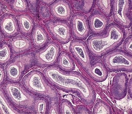
Image (×70) du champ encadré dans la figure 1.1B.

L’épididyme est composé d’un seul canal circonvolué dont on voit des coupes transversales (1), obliques (2) ou longitudinales (3). Le canal épididymaire est bordé par un épithélium, dont les détails sont indistincts. Au centre des canaux le matériel très coloré ou chromophile est formé des spermatozoïdes.

Le champ encadré est grossi 200 fois dans la figure 1.3.

Coloration: H–­­É
Grossissement: ×70


We'd very much appreciate receiving your comments and suggestions!We'd very much appreciate receiving your comments and suggestions!

Creative Commons Licence The text and images of this Histology Atlas, by Yves Clermont, Michael Lalli & Zsuzsanna Bencsath-Makkai, are licensed under a Creative Commons Attribution-Noncommercial-No Derivative Works 2.5 Canada Licence and cannot be modified without the written permission of the authors. Use of any text or images must carry an acknowledgement which includes a link to the original work.


HAweb/Zs B-M -> HAbase-generated on 2013-09-13