Contents Yves Clermont, Michael Lalli, Zsuzsanna Bencsath-Makkai
LIGHT-MICROSCOPIC HISTOLOGY ATLAS
     1. Epithelia
  1     2     3     4     5     6     7     8     9     10     11     12     13     14     15     16     17     18     19     20     21     22     23     24  

    1.5   Epididymis magnified 800 times Version française
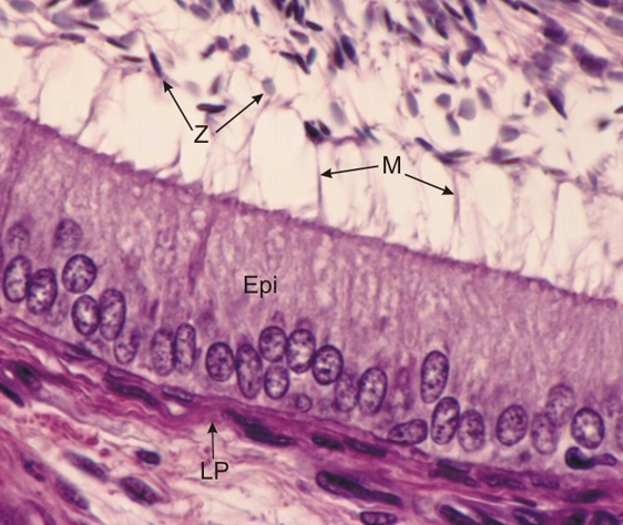
Higher-power view (×800) of the framed area of Figure 1.4.

The simple columnar epithelium (Epi) is composed of tall cells with their ovoid nuclei located at the base of the cytoplasm. The lateral cell walls are not visible owing to the oblique cut of the thick section. (See a thinner perpendicular section of the same epithelium in Figure 1.16).

The connective tissue or lamina propria (LP) under the epithelium, the nuclei of spermatozoa (Z) and the long flame-like clusters of microvilli (M) at the apex of the epithelial cells are also identified.

Stain: H–­­E
Magnification: ×800


We'd very much appreciate receiving your comments and suggestions!We'd very much appreciate receiving your comments and suggestions!

Creative Commons Licence The text and images of this Histology Atlas, by Yves Clermont, Michael Lalli & Zsuzsanna Bencsath-Makkai, are licensed under a Creative Commons Attribution-Noncommercial-No Derivative Works 2.5 Canada Licence and cannot be modified without the written permission of the authors. Use of any text or images must carry an acknowledgement which includes a link to the original work.


HAweb/Zs B-M -> HAbase-generated on 2013-09-13