Contents Yves Clermont, Michael Lalli, Zsuzsanna Bencsath-Makkai
LIGHT-MICROSCOPIC HISTOLOGY ATLAS
     1. Epithelia
  1     2     3     4     5     6     7     8     9     10     11     12     13     14     15     16     17     18     19     20     21     22     23     24  

    1.9   Urinary tubule lined with a simple squamous epithelium Version française
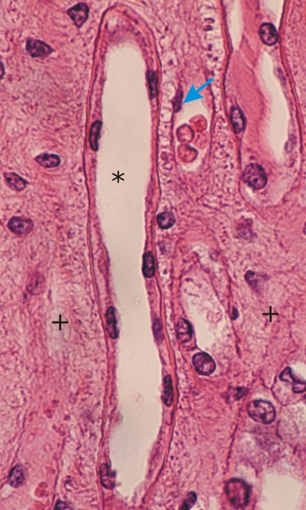
Longitudinal section of the thin limb of renal tubules showing a simple squamous epithelium (*). The flattened nuclei and cytoplasms of the squamous cells are evident but the lateral borderlines of the cells are not clear.

The nucleus of an endothelial cell (arrow) is visible in an adjacent small vessel containing a few red blood cells.

Connective tissue (+) is present between the urinary tubules and small vessels.

Stain: H–­­E
Magnification: ×600


We'd very much appreciate receiving your comments and suggestions!We'd very much appreciate receiving your comments and suggestions!

Creative Commons Licence The text and images of this Histology Atlas, by Yves Clermont, Michael Lalli & Zsuzsanna Bencsath-Makkai, are licensed under a Creative Commons Attribution-Noncommercial-No Derivative Works 2.5 Canada Licence and cannot be modified without the written permission of the authors. Use of any text or images must carry an acknowledgement which includes a link to the original work.


HAweb/Zs B-M -> HAbase-generated on 2013-09-13