|
||
| 1. Epithelia | ||
| 1 2 3 4 5 6 7 8 9 10 11 12 13 14 15 16 17 18 19 20 21 22 23 24 |
| |||
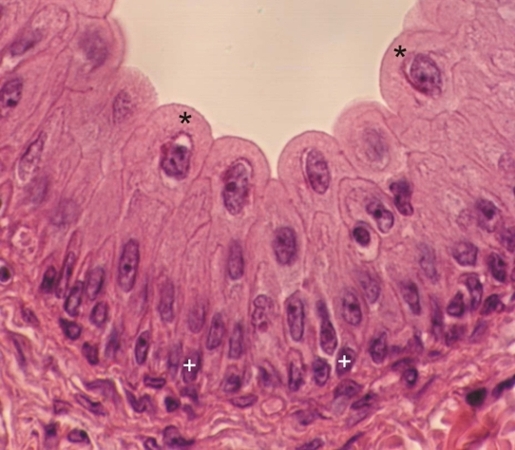 |
Pseudostratified epithelium of the urinary bladder is often described as being transitional because it continuously changes its morphology during the filling and emptying of the bladder.
In a filled bladder the epithelium is stratified and composed of only a few layers of squamous cells (not shown). In an empty bladder, as seen in this image, the stratified epithelium is composed of a layer of basal cells (+) applied to the lamina propria (LP). Superposed to these basal cells are vertical elongated cells of intermediate sizes. At the surface of the epithelium the cells (*) are voluminous and dome-shaped. Their spheroidal nucleus is large and occupies the centre of their abundant cytoplasm. A priori this epithelium appears as a stratified epithelium. In reality, as seen under the electron microscope, all the cells of this epithelium have processes reaching the basement membrane to which they tightly attach. Thus this epithelium should be considered pseudostratified. Stain: HE
|
||