Contents Yves Clermont, Michael Lalli, Zsuzsanna Bencsath-Makkai
LIGHT-MICROSCOPIC HISTOLOGY ATLAS
     1. Epithelia
  1     2     3     4     5     6     7     8     9     10     11     12     13     14     15     16     17     18     19     20     21     22     23     24  

    1.3   Epididymis magnified 200 times Version française
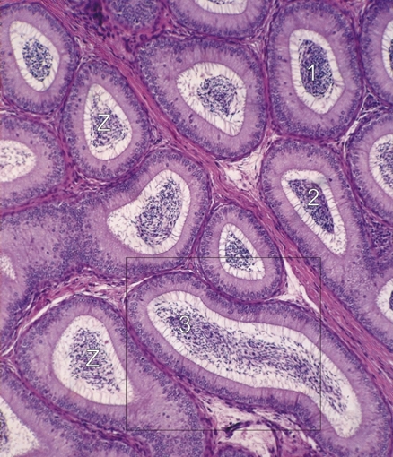
Higher-power view of the framed area in Figure 1.2 (×200).

Sections of the coiled epididymal duct are seen in cross (1), oblique (2) and longitudinal (3) sections.

In the simple columnar epithelium, the nuclei of the cells are seen at the base of the cytoplasm. The top or apex of the cells shows long filiform processes, which approach the clustered spermatozoa (Z).

The framed area is magnified 300 times in Figure 1.4.

Stain: H–­­E
Magnification: ×200


We'd very much appreciate receiving your comments and suggestions!We'd very much appreciate receiving your comments and suggestions!

Creative Commons Licence The text and images of this Histology Atlas, by Yves Clermont, Michael Lalli & Zsuzsanna Bencsath-Makkai, are licensed under a Creative Commons Attribution-Noncommercial-No Derivative Works 2.5 Canada Licence and cannot be modified without the written permission of the authors. Use of any text or images must carry an acknowledgement which includes a link to the original work.


HAweb/Zs B-M -> HAbase-generated on 2013-09-13