Contents Yves Clermont, Michael Lalli, Zsuzsanna Bencsath-Makkai
LIGHT-MICROSCOPIC HISTOLOGY ATLAS
     1. Epithelia
  1     2     3     4     5     6     7     8     9     10     11     12     13     14     15     16     17     18     19     20     21     22     23     24  

    1.16   Simple columnar stereociliated epithelium of the epididymal duct Version française
Simple columnar epithelium lining the lumen of the epididymal duct (see Figures 1.4 and 1.5).

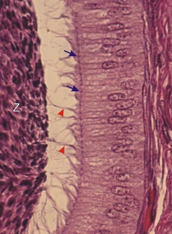
The lateral limits of the cytoplasm of epithelial cells are distinct. At the apex of these cells, perpendicular sections of the terminal bars, the sites of the junctional complexes, are clearly visible (arrows). At the apex of the cells there are long flame-like projections called stereocilia (red arrowheads). With the electron microscope, these stereocilia were shown to be composed of tufts of long non-motile microvilli.

The spermatozoa (Z), present in the lumen of the duct, are labelled.

Stain: H–­­E
Magnification: ×800


We'd very much appreciate receiving your comments and suggestions!We'd very much appreciate receiving your comments and suggestions!

Creative Commons Licence The text and images of this Histology Atlas, by Yves Clermont, Michael Lalli & Zsuzsanna Bencsath-Makkai, are licensed under a Creative Commons Attribution-Noncommercial-No Derivative Works 2.5 Canada Licence and cannot be modified without the written permission of the authors. Use of any text or images must carry an acknowledgement which includes a link to the original work.


HAweb/Zs B-M -> HAbase-generated on 2013-09-13