Contents Yves Clermont, Michael Lalli, Zsuzsanna Bencsath-Makkai
LIGHT-MICROSCOPIC HISTOLOGY ATLAS
     1. Epithelia
  1     2     3     4     5     6     7     8     9     10     11     12     13     14     15     16     17     18     19     20     21     22     23     24  

    1.1   Epididymis at low magnifications (×3 and ×6) Version française
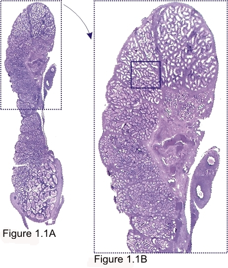
Epididymis of a dog.

Figure 1.1A (×3) is a longitudinal section of the whole epididymis.

Figure 1.1B (×6) is the framed area of Figure 1.1A. In these images the details of the convoluted epididymal duct that composes the organ are unclear.

The framed area of Figure 1.1B is magnified 70 times in Figure 1.2.

Stain: H–­­E
Magnification: ×3, ×6


We'd very much appreciate receiving your comments and suggestions!We'd very much appreciate receiving your comments and suggestions!

Creative Commons Licence The text and images of this Histology Atlas, by Yves Clermont, Michael Lalli & Zsuzsanna Bencsath-Makkai, are licensed under a Creative Commons Attribution-Noncommercial-No Derivative Works 2.5 Canada Licence and cannot be modified without the written permission of the authors. Use of any text or images must carry an acknowledgement which includes a link to the original work.


HAweb/Zs B-M -> HAbase-generated on 2013-09-13