Sommaire Yves Clermont, Michael Lalli, Zsuzsanna Bencsath-Makkai
ATLAS D'HISTOLOGIE EN MICROSCOPIE OPTIQUE
     1. Épithéliums
  1     2     3     4     5     6     7     8     9     10     11     12     13     14     15     16     17     18     19     20     21     22     23     24  
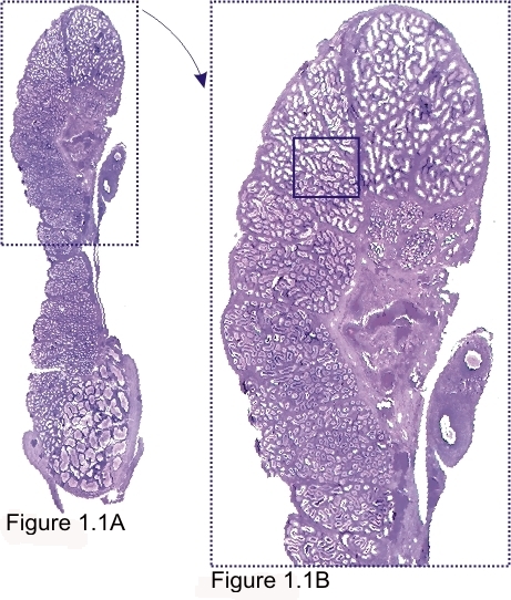
    1.1   L’épididyme de très faibles grossissements (×3 et ×6) English version
L’épididymis de chien.

La figure 1.1A (×3) est une coupe longitudinale de tout l’organe. À ce faible grossissement les détails histologiques de cet organe, formé d’un canal circonvolué, sont indistincts. La portion encadrée de figure 1.1A est grossie dans la figure 1B (×6).

La partie encadrée de la figure 1.1B est grossie 100 fois dans la figure 1.2.

Coloration: H–­­É
Grossissement: ×3, ×6


We'd very much appreciate receiving your comments and suggestions!We'd very much appreciate receiving your comments and suggestions!

Creative Commons Licence The text and images of this Histology Atlas, by Yves Clermont, Michael Lalli & Zsuzsanna Bencsath-Makkai, are licensed under a Creative Commons Attribution-Noncommercial-No Derivative Works 2.5 Canada Licence and cannot be modified without the written permission of the authors. Use of any text or images must carry an acknowledgement which includes a link to the original work.


HAweb/Zs B-M -> HAbase-generated on 2013-09-13