|
||
| 13. Urinary System | ||
| 1 2 3 4 5 6 7 8 9 10 11 12 13 14 15 16 17 18 19 20 21 22 23 24 25 | ||
| 26 27 |
| |||
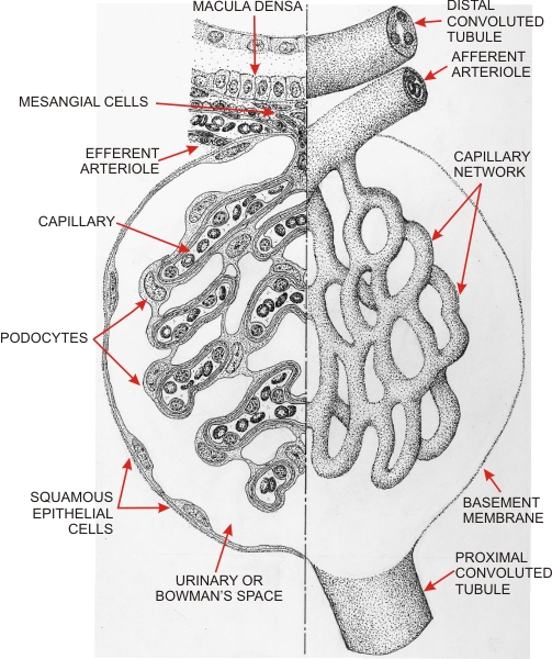 |
(Left) The drawing shows the components of the corpuscle as seen in a thin section. (Right) The drawing shows, in three dimensions, the afferent arteriole as it enters the corpuscle and branches into the capillary networks, or glomerulus. These capillaries unite at the vascular pole to form the efferent arteriole (shown in a longitudinal section, top left).
The connection of the corpuscle to a proximal convoluted tubule at the opposite pole of the corpuscle is schematically represented. At the vascular pole the thickened epithelium of the distal tubule forms the macula densa. This macula densa is located at the junction of the thick ascending limb of Henles loop and the distal convoluted tubule of the nephron (see Figure 13.5). Between the macula densa, the arterioles and the corpuscle there is a cushion of small connective cells called mesangial cells, some of which insert deeper into the corpuscle between the capillaries. In the section of the corpuscle on the left the following cells are represented:
The endothelial cells of capillaries, the podocytes and the basement membrane between these two layers constitute the site of formation of the urinary filtrate. The space between the parietal and the visceral epithelia is the urinary space also called Bowmans space. This space opens into the lumen of the proximal convoluted tubule.
|
||