|
||
| 13. Urinary System | ||
| 1 2 3 4 5 6 7 8 9 10 11 12 13 14 15 16 17 18 19 20 21 22 23 24 25 | ||
| 26 27 |
| |||
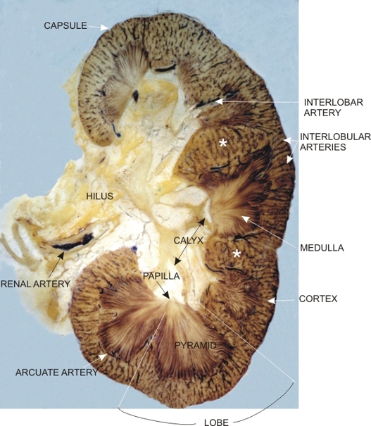 |
This is a slice of an unstained longitudinal section of a human kidney fixed and mounted between two plastic sheaths.
This longitudinal section passes through the hilus of the organ. The image shows the brownish kidney substance proper, or parenchyma, with its cortex and medulla. (Compare this image with the drawing in Figure 13.2.) In the hilus, the slightly denser tissue in the yellowish fat is the wall of the calyces. In the kidney substance proper, the cortex is streaked with fine brown lines corresponding to cortical radial vessels. The finely striated medulla corresponds to the pyramids with their apixes or papillae directed toward the lumina of the calyces. The renal columns (*) are extensions of the cortex between the pyramids that reach the hilus of the kidney. A segment of the renal artery is visible at the entrance of the hilus and a few oblique or cross sections of large arteries are seen in the kidney itself. The limits of a lobe are shown at the bottom of the image.
|
||