Contents Yves Clermont, Michael Lalli, Zsuzsanna Bencsath-Makkai
LIGHT-MICROSCOPIC HISTOLOGY ATLAS
     13. Urinary System
  1     2     3     4     5     6     7     8     9     10     11     12     13     14     15     16     17     18     19     20     21     22     23     24     25  
  26     27  

    13.27   Urinary bladder Version française
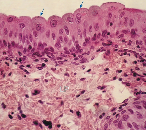
Mucosa of the urinary bladder.

At the surface of this mucosa, the epithelium, classified as transitional, is composed of several layers of cells.

The superficial epithelial cells are large and dome-shaped (arrows). All the cells of this epithelium become squamous in distended bladders. Thus, this epithelium continually varies in thickness.

This epithelium is sitting on a lamina propria composed of dense connective tissue.

Stain: H–­­E
Magnification: ×600


We'd very much appreciate receiving your comments and suggestions!We'd very much appreciate receiving your comments and suggestions!

Creative Commons Licence The text and images of this Histology Atlas, by Yves Clermont, Michael Lalli & Zsuzsanna Bencsath-Makkai, are licensed under a Creative Commons Attribution-Noncommercial-No Derivative Works 2.5 Canada Licence and cannot be modified without the written permission of the authors. Use of any text or images must carry an acknowledgement which includes a link to the original work.


HAweb/Zs B-M -> HAbase-generated on 2013-09-13