Contents Yves Clermont, Michael Lalli, Zsuzsanna Bencsath-Makkai
LIGHT-MICROSCOPIC HISTOLOGY ATLAS
     13. Urinary System
  1     2     3     4     5     6     7     8     9     10     11     12     13     14     15     16     17     18     19     20     21     22     23     24     25  
  26     27  

    13.11   Renal corpuscle Version française
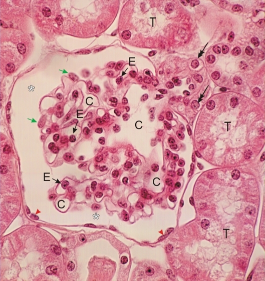
Thin section of a renal corpuscle with an arteriole connected to the capillaries (C) of a glomerular tuft.

The large cells forming the wall of this arteriole (double-headed arrows) are modified smooth muscle cells called juxtaglomerular cells. They are endocrine cells that secrete renin. Along with the macula densa and mesangial cells, they form the juxtaglomerular apparatus.

The other cells of the glomerulus and of the Bowman’s capsule are also labelled: endothelial nuclei (E) of capillaries, podocytes (green arrows) and cells of the parietal layer (arrowheads). The Bowman’s space (*) and the renal tubules (T) surrounding the renal corpuscle are also identified.

Stain: H–­­E
Magnification: ×600


We'd very much appreciate receiving your comments and suggestions!We'd very much appreciate receiving your comments and suggestions!

Creative Commons Licence The text and images of this Histology Atlas, by Yves Clermont, Michael Lalli & Zsuzsanna Bencsath-Makkai, are licensed under a Creative Commons Attribution-Noncommercial-No Derivative Works 2.5 Canada Licence and cannot be modified without the written permission of the authors. Use of any text or images must carry an acknowledgement which includes a link to the original work.


HAweb/Zs B-M -> HAbase-generated on 2013-09-13