Contents Yves Clermont, Michael Lalli, Zsuzsanna Bencsath-Makkai
LIGHT-MICROSCOPIC HISTOLOGY ATLAS
     13. Urinary System
  1     2     3     4     5     6     7     8     9     10     11     12     13     14     15     16     17     18     19     20     21     22     23     24     25  
  26     27  

    13.9   Renal corpuscle Version française
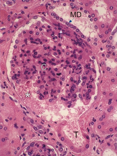
Section of a renal corpuscle.

This field shows the glomerular tuft of the corpuscle (C) attached to a cushion of mesangial cells (arrow) next to the macula densa (MD).

The glomerular tuft is suspended in the urinary space filled with precipitated proteins (*).

Facing the glomerular tuft and opposite the vascular pole, the urinary space is continuous with the lumen of the proximal convoluted tubule (T) of the nephron.

Stain: H–­­E
Magnification: ×700


We'd very much appreciate receiving your comments and suggestions!We'd very much appreciate receiving your comments and suggestions!

Creative Commons Licence The text and images of this Histology Atlas, by Yves Clermont, Michael Lalli & Zsuzsanna Bencsath-Makkai, are licensed under a Creative Commons Attribution-Noncommercial-No Derivative Works 2.5 Canada Licence and cannot be modified without the written permission of the authors. Use of any text or images must carry an acknowledgement which includes a link to the original work.


HAweb/Zs B-M -> HAbase-generated on 2013-09-13