Contents Yves Clermont, Michael Lalli, Zsuzsanna Bencsath-Makkai
LIGHT-MICROSCOPIC HISTOLOGY ATLAS
     13. Urinary System
  1     2     3     4     5     6     7     8     9     10     11     12     13     14     15     16     17     18     19     20     21     22     23     24     25  
  26     27  

    13.10   Renal corpuscle Version française
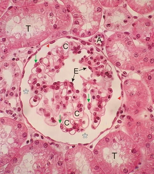
Thin section of a renal corpuscle.

A cross section of an arteriole (A) is seen proximal to the vascular pole of the corpuscle.

The glomerular tuft shows the capillaries (C) and some nuclei of the endothelial cells (E).

The larger nuclei and cytoplasm of podocytes (arrows) are easily identifiable in this preparation.

The squamous epithelial cells forming the parietal layer of Bowman’s capsule (arrowheads) and the urinary space (*) are indicated.

Sections of proximal convoluted tubules (T) surround the corpuscle.

Stain: H–­­E
Magnification: ×500


We'd very much appreciate receiving your comments and suggestions!We'd very much appreciate receiving your comments and suggestions!

Creative Commons Licence The text and images of this Histology Atlas, by Yves Clermont, Michael Lalli & Zsuzsanna Bencsath-Makkai, are licensed under a Creative Commons Attribution-Noncommercial-No Derivative Works 2.5 Canada Licence and cannot be modified without the written permission of the authors. Use of any text or images must carry an acknowledgement which includes a link to the original work.


HAweb/Zs B-M -> HAbase-generated on 2013-09-13