|
||
| 13. Urinary System | ||
| 1 2 3 4 5 6 7 8 9 10 11 12 13 14 15 16 17 18 19 20 21 22 23 24 25 | ||
| 26 27 |
| |||
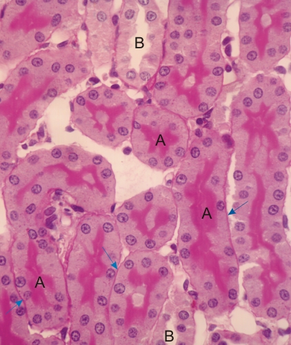 |
Cortex of a rodent kidney stained with PAS.
This field shows sections of several proximal convoluted tubules (A) and of a few distal convoluted tubules (B). The brush border of the proximal convoluted tubules is PAS-positive, owing to the presence of glycoproteins associated with the plasma membrane of the microvilli. The thin basement membrane of these tubules is also PAS-positive (blue arrows). It is noticeable that the apex of the epithelial cells of the distal convoluted tubules (B) is not stained with PAS. Stain: PAS-Hematoxylin
|
||