|
||
| 13. Urinary System | ||
| 1 2 3 4 5 6 7 8 9 10 11 12 13 14 15 16 17 18 19 20 21 22 23 24 25 | ||
| 26 27 |
| |||
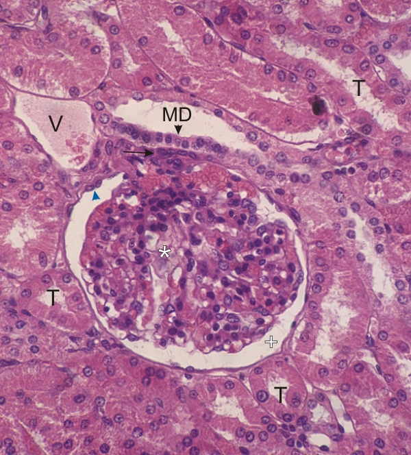 |
Section of a renal corpuscle surrounded by proximal convoluted tubules (T).
The plane of section of the corpuscle crosses the vascular pole of the corpuscle and shows the macula densa (MD). The macula densa is formed of closely packed cuboidal epithelial cells lining the terminal end of the thick ascending limb of Henles loop. It is attached to a collection of mesangial cells (arrow) connected to the glomerular tuft (*) (see Figure 13.8). The glomerular tuft is composed of a network of capillaries or glomerulus, and of podocytes which form the visceral layer of Bowmans capsule. The podocytes are difficult to differentiate from endothelial cells of capillaries in this section. The glomerular tuft in located in the urinary space also called Bowmans space (+), which is delimited externally by the parietal layer of Bowmans capsule (arrowhead). A venule is also labelled (V). Stain: HE
|
||