Contents Yves Clermont, Michael Lalli, Zsuzsanna Bencsath-Makkai
LIGHT-MICROSCOPIC HISTOLOGY ATLAS
     13. Urinary System
  1     2     3     4     5     6     7     8     9     10     11     12     13     14     15     16     17     18     19     20     21     22     23     24     25  
  26     27  

    13.6   Kidney cortex Version française
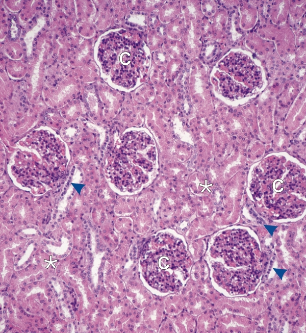
Section of the kidney cortex showing several renal corpuscles (C).

Several of the renal corpuscles show, at their vascular poles, the epithelial thickening, or macula densa (arrowheads) of the thick ascending limbs of Henle’s loops (see Figure 13.5).

The surrounding tubular profiles (*) belong to many proximal convoluted tubules and to some distal convoluted tubules of the nephrons.

Stain: H–­­E
Magnification: ×150


We'd very much appreciate receiving your comments and suggestions!We'd very much appreciate receiving your comments and suggestions!

Creative Commons Licence The text and images of this Histology Atlas, by Yves Clermont, Michael Lalli & Zsuzsanna Bencsath-Makkai, are licensed under a Creative Commons Attribution-Noncommercial-No Derivative Works 2.5 Canada Licence and cannot be modified without the written permission of the authors. Use of any text or images must carry an acknowledgement which includes a link to the original work.


HAweb/Zs B-M -> HAbase-generated on 2013-09-13