 |
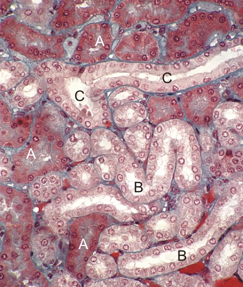
Section of the kidney cortex stained with Massons trichrome.
This field shows sections of three types of tubules:
- The acidophilic proximal convoluted tubules of the nephron are deeply stained.
- The distal convoluted tubules of the nephron have larger lumina and their epithelial cells are lightly stained and striated but their lateral cytoplasmic membranes are indistinct.
- The collecting ducts show epithelial cells with lightly stained non-striated cytoplasms, but with clear lateral cell membranes.
Table I. Identification criteria for tubules and ducts in the
kidney cortex |
| Cell types |
Staining |
Cell apex* |
Lateral cell wall** |
| Proximal convoluted tubules |
Strongly acidophilic |
Large brush border |
Indistinct |
| Distal convoluted tubules |
Lightly acidophilic |
Brush border less prominent |
Indistinct |
| Collecting duct |
Pale |
Absent brush border |
Distinct |
*All cells show apical microvilli. In the case of "brush borders," the microvilli are long and abundant. In the case of "invisible brush borders," the microvilli are small and sparse.
**The lateral cell walls are "distinct" when the cytoplasmic membranes are clearly visible from the base to the apex of the epithelial cells. The lateral cell walls are "indistinct" because the plasma membranes present numerous lateral projections and are thus not visible with the light microscope.
|
Stain: Massons Trichrome
Magnification: ×400
|