Contents Yves Clermont, Michael Lalli, Zsuzsanna Bencsath-Makkai
LIGHT-MICROSCOPIC HISTOLOGY ATLAS
     13. Urinary System
  1     2     3     4     5     6     7     8     9     10     11     12     13     14     15     16     17     18     19     20     21     22     23     24     25  
  26     27  

    13.19   Kidney: Outer medulla Version française
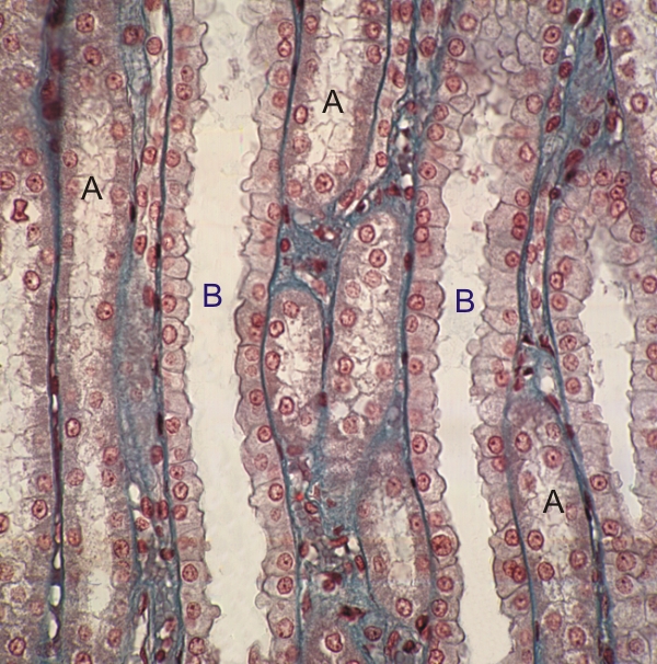
Outer medulla of a kidney stained with Masson’s trichrome.

This field shows longitudinal sections of thick descending or ascending limbs of Henle’s loops. Their acidophilic epithelial cells have indistinct lateral cytoplasmic membranes (A).

The large collecting ducts are lined with cuboidal cells which show distinct lateral cytoplasmic membranes (B).

The intertubular connective tissue is stained green.

Stain: Masson’s Trichrome
Magnification: ×400


We'd very much appreciate receiving your comments and suggestions!We'd very much appreciate receiving your comments and suggestions!

Creative Commons Licence The text and images of this Histology Atlas, by Yves Clermont, Michael Lalli & Zsuzsanna Bencsath-Makkai, are licensed under a Creative Commons Attribution-Noncommercial-No Derivative Works 2.5 Canada Licence and cannot be modified without the written permission of the authors. Use of any text or images must carry an acknowledgement which includes a link to the original work.


HAweb/Zs B-M -> HAbase-generated on 2013-09-13