|
||
| 13. Urinary System | ||
| 1 2 3 4 5 6 7 8 9 10 11 12 13 14 15 16 17 18 19 20 21 22 23 24 25 | ||
| 26 27 |
| |||
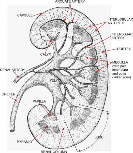 |
Diagram of a human kidney which corresponds to the thin slice of the kidney shown in Figure 13.1. (View Figure 13.1 and Figure 13.2 side by side)
The drawing shows the hilus in the concavity of the organ, along with the kidney parenchyma. The ureter enters the hilus and is continuous with the renal pelvis. It branches into calyces which take a funnel or cup shape at their extremities. These cups are applied to the papillae of the pyramids. Between the calyces and along the pelvis are large arteries and accompanying veins (these veins are not shown here). The kidney parenchyma is subdivided into two main zones: the cortex underlying the capsule and the medulla, located deeper and next to the hilus. The kidney tissue is organized into lobes (from 8 to 18 per kidney in humans). Each lobe is composed of a pyramid surrounded by the cortex which extends deeper as renal columns. The lateral boundaries of the lobes are not well demarcated but cross the mid-portions of the renal columns, as indicated on the diagram by the two lines delimiting the lobe (bottom right). The striated pyramids show two zones: a dark outer medulla and a pale inner medulla. The intra-renal branches of renal arteries are schematically represented (top). The renal arteries of the hilus branch out and give rise to the interlobar arteries which enter the kidney parenchyma at the level of the renal columns. They then turn 90 degrees to form arches, the arcuate arteries, running along the borderline of the cortex and medulla. Numerous smaller radial arteries, arising from the arcuate arteries, are oriented toward the capsule. These radial arterioles branch laterally into arterioles that reach the renal corpuscles (not shown here but illustrated in Figure 13.5).
|
||