Contents Yves Clermont, Michael Lalli, Zsuzsanna Bencsath-Makkai
LIGHT-MICROSCOPIC HISTOLOGY ATLAS
     13. Urinary System
  1     2     3     4     5     6     7     8     9     10     11     12     13     14     15     16     17     18     19     20     21     22     23     24     25  
  26     27  

    13.4   Kidney cortex Version française
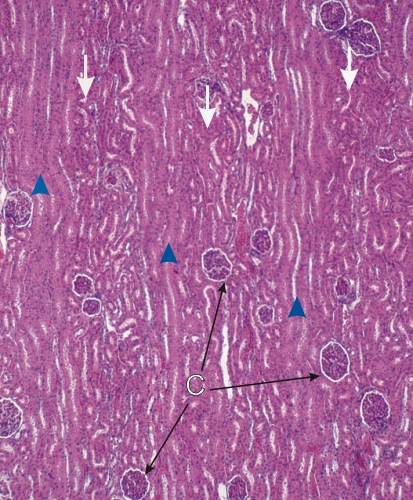
Section of the kidney cortex.

The spheroidal renal corpuscles (C) are easily identified.

The rest of the tissue is composed of tubules that belong to nephrons, some of which are convoluted (arrows), while others are straight and form parallel fascicles (arrowheads). These fascicles are called medullary rays although they are located in the cortex but are in continuity with the medulla.

The details of the various types of urinary tubules, not seen at this low magnification, will be examined at higher magnifications in subsequent images.

Stain: H–­­E
Magnification: ×50


We'd very much appreciate receiving your comments and suggestions!We'd very much appreciate receiving your comments and suggestions!

Creative Commons Licence The text and images of this Histology Atlas, by Yves Clermont, Michael Lalli & Zsuzsanna Bencsath-Makkai, are licensed under a Creative Commons Attribution-Noncommercial-No Derivative Works 2.5 Canada Licence and cannot be modified without the written permission of the authors. Use of any text or images must carry an acknowledgement which includes a link to the original work.


HAweb/Zs B-M -> HAbase-generated on 2013-09-13