Contents Yves Clermont, Michael Lalli, Zsuzsanna Bencsath-Makkai
LIGHT-MICROSCOPIC HISTOLOGY ATLAS
     13. Urinary System
  1     2     3     4     5     6     7     8     9     10     11     12     13     14     15     16     17     18     19     20     21     22     23     24     25  
  26     27  

    13.14   Renal cortex Version française
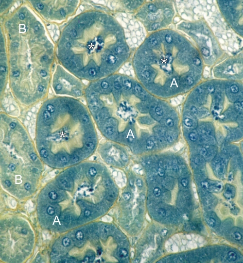
Cortex of a rodent kidney stained with TPA.

The proximal convoluted tubules (A) have epithelial cells with a heavily stained cytoplasm and a distinct apical yellowish brush border. In the stellate lumina the blue granular material (*) corresponds to precipitated proteins.

Some segments of the proximal convoluted tubules (B) are less chromophilic. This indicates that proximal convoluted tubules have more than one subsegment.

Stain: TPA
Magnification: ×600


We'd very much appreciate receiving your comments and suggestions!We'd very much appreciate receiving your comments and suggestions!

Creative Commons Licence The text and images of this Histology Atlas, by Yves Clermont, Michael Lalli & Zsuzsanna Bencsath-Makkai, are licensed under a Creative Commons Attribution-Noncommercial-No Derivative Works 2.5 Canada Licence and cannot be modified without the written permission of the authors. Use of any text or images must carry an acknowledgement which includes a link to the original work.


HAweb/Zs B-M -> HAbase-generated on 2013-09-13