Contents Yves Clermont, Michael Lalli, Zsuzsanna Bencsath-Makkai
LIGHT-MICROSCOPIC HISTOLOGY ATLAS
     13. Urinary System
  1     2     3     4     5     6     7     8     9     10     11     12     13     14     15     16     17     18     19     20     21     22     23     24     25  
  26     27  

    13.24   Tip of a renal pyramid Version française
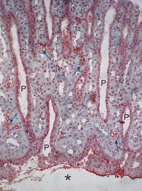
Longitudinal section through the tip a renal pyramid of a dog.

This field shows large collecting ducts also called papillary ducts (P).

Some of these ducts approach or connect to the calyx (*).

Several small vessels (arrows), some containing red blood cells, are seen in the connective tissue.

Stain: Masson’s Trichrome
Magnification: ×150


We'd very much appreciate receiving your comments and suggestions!We'd very much appreciate receiving your comments and suggestions!

Creative Commons Licence The text and images of this Histology Atlas, by Yves Clermont, Michael Lalli & Zsuzsanna Bencsath-Makkai, are licensed under a Creative Commons Attribution-Noncommercial-No Derivative Works 2.5 Canada Licence and cannot be modified without the written permission of the authors. Use of any text or images must carry an acknowledgement which includes a link to the original work.


HAweb/Zs B-M -> HAbase-generated on 2013-09-13