|
||
| 2. Connective Tissue | ||
| 1 2 3 4 5 6 7 8 9 10 11 12 13 14 15 16 17 18 19 |
| |||
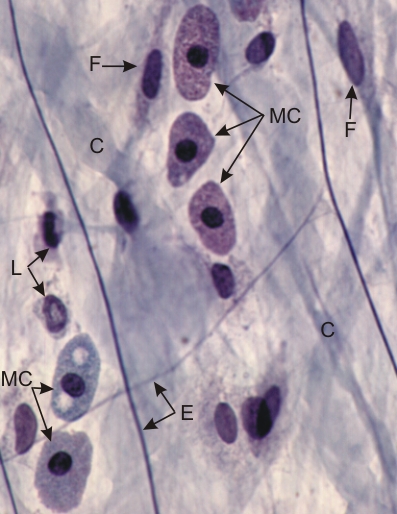 |
Whole mount of a rat mesentery stained with Massons trichrome and iron hematoxylin.
In this loose connective tissue the three granulated mast cells above (MC) (top, centre) show a small dark spherical nucleus in abundant cytoplasm filled with granules. Two mast cells (bottom, left) are degranulated. These cells secrete heparin, an anticoagulant. The following elements are also labelled: fibrocytes (F), leukocytes (L), elastic fibres (E) and collagen fibres (C). The nuclei of mesothelial cells are not present in this preparation. Stain: Massons Trichrome
|
||