|
||
| 2. Connective Tissue | ||
| 1 2 3 4 5 6 7 8 9 10 11 12 13 14 15 16 17 18 19 |
| |||
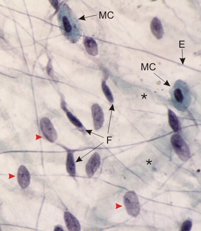 |
Whole mount of a rat mesentery stained with Massons trichrome and iron hematoxylin.
A small piece of mesentery was dissected and deposited on a glass slide and fixed. Following staining, this thin layer of tissue was covered with a droplet of resin and a thin glass cover slip. In such a preparation the cells are not sectioned with a microtome and are complete between the glass slide and the cover slip. Some elastic fibres (E) are stained blue with iron hematoxylin and the collagen (*) appears as pale wavy greyish bands. A few fibrocytes (F), with their extended cytoplasmic processes, and two degranulated mast cells (MC) are labelled. The cells, with large flat nuclei (arrowheads) and indistinct cytoplasmic membrane limits, belong to the simple squamous epithelial cells (or mesothelium) covering the surfaces of the mesentery. Stain: Massons Trichrome
|
||