|
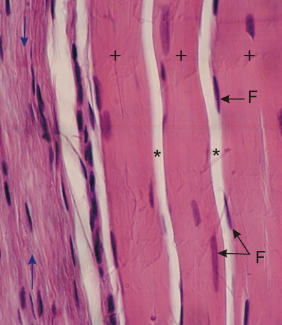
Dense connective tissue forming a tendon (right) adjacent to a less dense layer of connective tissue (left).
In the tendon the three collagen fibres (+) are composed of closely packed bundles of type I collagen fibrils parallel to each other. Flattened fibrocytes (F) are rare. The unstained spaces between these collagen fibres are fixation artefacts (*).
Adjacent to the tendon (left), the bundles of type I collagen fibrils appear smaller but are also parallel to each other (arrows). Fibrocytes are more numerous in the less dense layer of connective tissue.
Stain: HE
Magnification: ×700
|