|
||
| 2. Connective Tissue | ||
| 1 2 3 4 5 6 7 8 9 10 11 12 13 14 15 16 17 18 19 |
| |||
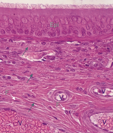 |
Section of the tracheal mucosa stained with hematoxylin-eosin.
This image shows the dense connective tissue underlying the ciliated epithelium (Epi). This connective tissue is composed mainly of intertwined bundles of acidophilic type I collagen fibres (arrows). Elastic fibres are also present in the tissue but cannot be distinguished from collagen (see Figure 2.4). The nuclei of fibrocytes (F) and small vessels (V) are labelled. Stain: HE
|
||