|
||
| 2. Connective Tissue | ||
| 1 2 3 4 5 6 7 8 9 10 11 12 13 14 15 16 17 18 19 |
| |||
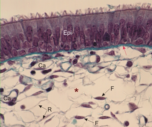 |
Section of the mucosa of the larynx stained with Massons trichrome.
In this field the connective tissue is loose and shows a few fibres. Some small collagen fibrils or reticular fibres (R) are seen around empty-looking spaces (*). The main connective tissue cells are the fibrocytes (F) showing fusiform nuclei and a scanty cytoplasm. Underlying the ciliated epithelium (Epi) is a relatively thick layer stained green (red arrow) composed of several extracellular elements that cannot be distinguished from one another, i.e. a thin basement membrane (composed of type IV collagen, proteoglycans, laminin and fibronectin), anchoring fibrils (type VII collagen), reticular fibrils (type III collagen), microfibrils (fibrilin), and type I collagen fibrils. Some capillaries (Ca) are indicated. Stain: Massons Trichrome
|
||