Contents Yves Clermont, Michael Lalli, Zsuzsanna Bencsath-Makkai
LIGHT-MICROSCOPIC HISTOLOGY ATLAS
     2. Connective Tissue
  1     2     3     4     5     6     7     8     9     10     11     12     13     14     15     16     17     18     19  

    2.9   Connective tissue Version française
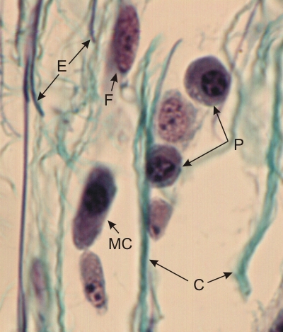
Section of the connective tissue of a tongue stained with Masson’s trichrome and iron hematoxylin.

In addition to a few elastic fibres stained blue (E) with iron hematoxylin, some small collagen fibres (C) are stained green with Masson’s trichrome.

In this field the following cells are identified: a fibrocyte (F), a mast cell (MC) and two plasma cells (P). These plasma cells, which derive from lymphocytes, secrete antibodies during antigenic reactions.

Stain: Masson’s Trichrome
Magnification: ×900


We'd very much appreciate receiving your comments and suggestions!We'd very much appreciate receiving your comments and suggestions!

Creative Commons Licence The text and images of this Histology Atlas, by Yves Clermont, Michael Lalli & Zsuzsanna Bencsath-Makkai, are licensed under a Creative Commons Attribution-Noncommercial-No Derivative Works 2.5 Canada Licence and cannot be modified without the written permission of the authors. Use of any text or images must carry an acknowledgement which includes a link to the original work.


HAweb/Zs B-M -> HAbase-generated on 2013-09-13